Friday, April 30, 2010

Why the Outer Banks will rock your face off

At 5:00 a.m., I was hanging out in the parking lot with my Marine Biology lab notebook and camping gear. I stood there waiting for the rest of my BIO 412 classmates to show up, trying not to move too much because it made the spinning in my head worse. (I knew I would probably be sleeping most of the van ride to North Carolina, so I had gone out and gotten my drink on.) I shivered, wondering vaguely how long it would take for AU Safety Services to investigate a report of a drunkard camping out in the parking lot, when other students began to arrive, yawning and lugging camping gear.  Once all the gear was loaded in the vans, Doctors Posner and Saunders, two of the AU Biology department faculty, began the drive.  We were on our way!

Between being subjected to Tom’s awful taste in music approximately seven thousand fifty six times, I managed to take some awesome pictures of our trip. Enjoy!

Camp Awesome: We came here for taxon identification, sleep, and snackage.
At 11:00 p.m. the last night, 30mph gusts came out of nowhere and ripped our tents down.  The poles of two tents were left snapped and useless.

Even 15-foot tall sand dunes are futile attempts to prevent the erosion that barrier islands experience naturally.


We went kayaking in a salt marsh and I spotted a duck. I was able to get close enough to take this sweet pic right as it took off.


The Cape Hatteras Light Station. It is 198 feet tall, and you must climb 257 steps to reach the top!


The view from the top was insane! It took me three minutes just to stop hugging the wall and stand by the edge to take this picture.
The entire climb up, I had a deathgrip on the railing while trying to look like I wasn’t having a claustrophobic / heights freakout.


The Littoraria snails will climb the Spartina marsh grass to escape predation by crabs below. They can hold on even as they sleep by secreting a sticky substance onto the spot and just hang there, sleeping.


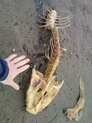
We came across this huge fish skeleton in the salt marsh. I took a picture with my hand next to it to set the scale for how huge this thing was.
We wanted such a complete specimen for our lab back in Ohio, so we wrapped it in trash bags and stored it in our lunchmeat cooler.


Once you get the chance, go check out the Outer Banks! But whatever you do, don’t feed the Laughing gulls! They will stalk you forever if you do.

Wednesday, April 28, 2010

Shower Anyone??


For anyone that has taken a microbiology course, especially here at Ashland you feel completely dirty after finishing up experiments in lab. So what better way of cleansing yourself from a hard days work in the lab, experimenting with bacteria that you are trying to identify for your 25 page lab report. Who doesn’t like waking up and taking a hot shower before school or work to feel clean throughout the day? What people don’t know is that the showerhead is a perfect environment for microbes. It’s moist, warm, dark and frequently replenished with low amounts of nutrients for these microbes to feed on. Who could have thought that these microbes can lead to pulmonary disease and other health risks such as asthma and bronchitis? Believe it or not it is true and studies have proven that microbes do exist.

The common microbe that was found in high levels was Mycobacterium avium, a pathogen that is linked to pulmonary disease. M. avium and related pathogens were seen clumped together on showerheads in slimy biofilms. Studies shown that the showerheads were more than 100 times the background levels of municipal water when compared to the showerhead when they were taken off. "If you are getting a face full of water when you first turn your shower on, that means you are probably getting a particularly high load of Mycobacterium avium, which may not be too healthy," said study leader Norman Pace.


These biofilms were swabbed on interior surfaces of 45 showerheads from nine cities in the United States. Researchers found that nearly a third of the showerheads tested were harboring these pathogens. While it is rarely a problem for most healthy people, those with weakened immune systems, like the elderly, pregnant women or those who are fighting off other diseases, can be susceptible to infection.

One showerhead in the study was found with high loads of the pathogen Mycobacterium gordonae. The showerhead was cleaned with a bleach solution, but later tests on the showerhead showed the bleach treatment had actually caused a three-fold increase in M. gordonae, indicating a general resistance of mycobacteria species to chlorine.
Research at National Jewish Hospital in Denver indicates that increases in lung infections in the United States in recent decades from so-called “non-tuberculosis” mycobacteria species like M. avium may be linked to people taking more showers and few¬er baths, said Pace. Water spurting from showerheads can distribute pathogen-filled droplets that float in the air and can easily be inhaled into the deepest parts of the lungs.


So what should I do about taking showers? I really don’t want to be the smelly kid in class that everyone talks about. Well, fellow readers you can always replace the showerhead. Research shows that that plastic showerheads allow for more bacteria to clump together when compared to the metal showerhead counterpart. You can also allow the water to run at the hottest level for a couple of minutes and then turn the water to a tolerable level. Not welling to give up that 1980’s showerhead? Solution, take a bath.

Falkinham III, Joseph, and Michael Iseman. "Mycobacterium avium in a shower linked to pulmonary disease." Journal of Water and Health. 6.2 (2008): 209-11. Print.

Feazel, Leah, Laura Baumgartner, and Kristin Peterson. "Opportunistic pathogens enriched in showerhead biofilms." Proceedings of the National Academy of Sciences of the United States of America 12.2 (2009): n. pag. Web. 28 Apr 2010.

Can sleep deficiency really kill you?

As I have stated before I am one of the unfortunate 64 million people in America that suffer from insomnia. My dad has it, my brother has it, and I have it. Without insomnia I probably wouldn’t get a whole lot of things done (like this blog post). Fortunately for me having insomnia also has some other perks, such as getting to watch TV shows about insomnia. Earlier tonight I watched a show on the National Geographic channel about a form of insomnia that I was completely unaware of and I am sure I am not the only person who is unaware of it since it affects only 40 families in the entire world. This type of insomnia is called Fatal Familial Insomnia or FFI. I never thought that Insomnia could kill someone, but this form of Insomnia makes every person who is diagnosed with it a victim.

I thought that I would go over the sleep cycle a little bit first to help you understand how sleeping works but since a colleague has already posted a blog about it I will just set up a link to that post here http://fourthirstypandas.blogspot.com/2010/04/sweet-dream-or-beautiful-nightmare.html.

FFI has been characterized as a genetic disorder caused by a mutation at codon 178 of the prion protein gene. This is also a problem in the condition called “Mad Cow Disease.” Prions are proteins that attack the nervous system and cause the symptoms associated with “Mad Cow Disease.” In FFI the cause of the prion protein is genetic, where a single base pair is coded incorrectly. This is one of 3 billion base pairs known in the human genome. In FFI the prions accumulate in the Thalamus in the brain. The Thalamus was never thought to control sleep, but it does transmit signals to the cortex of the brain. In patients with FFI lesions occurred in the Thalamus and the Cortex of the brain. With FFI 90% of the neurons in the Thalamus have disappeared.


ally sleep has been one of the hardest things to study, but some recent studies using a PET scan and a tagged amino acid may give us some clues as to why we need to sleep. One of the theories is that we need sleep to repair proteins in the cells of the brain, which can’t happen while we are awake because the brain is too busy and has too many processes happening. During a normal day while we are awake we accumulate adenosine in our brain, which signals our bodies to sleep. Sleep would eliminate the adenosine and produce more proteins for repair of the brain cells.

Humans have varying levels of sleep, but so do many animals in nature. A normal human gets somewhere between 7 and 8 hours of sleep per night, but an animal such as a lion gets up to 15 hours a day. A elephant on the other hand gets around 4 hours of sleep. One of the theories about this is that predator species can sleep more because they don’t generally have many predators, while the prey species get much less sleep so they can evade their predators. But what if we could sleep and be awake at the same time. I know this sound ridiculous, but a few animals in nature already have achieved this process. If we could somehow find out how to translate this to humans we could eliminate FFI and other sleep associated disorders.

One of the species in nature that sleeps all the time but we never see it are dolphins. I always think of a dolphin as an animal that is always moving and swimming. A study done in San Diego evaluated if a dolphin lost any mental abilities when forced to stay alert for multiple days. The dolphin was trained to detect a swimmer that was in the bay while the dolphin was detained in a fenced area. When the dolphin detected the swimmer it hit a switch on the dock. The dolphin showed no decline in activity and detected every time the swimmer was in the bay. The dolphin slept throughout the entire experiment. This is possible due to dolphins having something called unihemispheric sleep. Throughout the dolphins life one half of their brain is active while the other is sleeping and then they switch. Another animal that is theorized to do this is many different avian species. Studies have shown that they can sleep while also watching for predators while they are on land, but it is still unclear wheather they can sleep while flying.

So far for all terrestrial mammals, sleep is needed. After several days of sleep deprivation there is a drastic decrease in overall health. One side effect is diabetes, due to insulin resistance that is accumulated. There is also a decrease in lymphocytes which fight bacterial infections in the body. In most mammals if sleep is deprived for 2 weeks death will occur. In patients with FFI death usually occurs from 7 to 36 months, and the unfortunate thing is that once symptoms of FFI start they never go away until the patient dies. The unfortunate thing about this disease is that there is currently no cure. And if one family member is ever diagnosed with FFI there is a 50% chance that their children will be diagnosed with it as well since it is a dominant gene.

So now that I have accumulated a massive amount of adenosine in my brain I think I will go sleep and I encourage everybody else to get sufficient amounts of sleep as well.

References:

1. National Geographic Explorer: “Fatal Insomnia”, aired April 27, 2010

2. Fatal familial insomnia: clinical features and molecular genetics; PIETRO CORTELLI, PIERLUIGI GAMBETTI, PASQUALE MONTAGNA and ELIO LUGARESI; J. Sleep Res. (1999) 8, Suppl. 1, 23-29; European Sleep Research Society

3. Montagna P, Gambetti P, Cortelli P, Lugaresi E (2003). "Familial and sporadic fatal insomnia". Lancet Neurol 2 (3): 167–76. doi:10.1016/S1474-4422(03)00323-5

4. Almer G, Hainfellner JA, Brücke T, et al. (1999). "Fatal familial insomnia: a new Austrian family". Brain 122 ( Pt 1): 5–16. doi:10.1093/brain/122.1.5.

Tuesday, April 27, 2010

Are tanning beds a cancer risk?

Being the scrawny white nerd that I am, I don’t tan… I burn. This concerned me greatly since I would soon be traveling to the Outer Banks in my Marine Biology class. I wanted to go swimming in the ocean, but that would mean my un-tanned, blinding white-ness would be on display. Not wanting to look completely ridiculous, I contemplated going tanning for the first time ever.


But I was worried. Tanning lotions were out because I don’t like the thought of smearing skin-altering chemicals on myself. And I had heard from many sources that tanning beds cause cancer. So I decided to look into the matter.

According to this study I found [1], exposure to tanning beds actually does increase the risk of developing malignant melanoma (skin cancer).

Where are they getting that from?

Ting and his crew wanted to test the hypothesis that increased exposure to tanning beds was linked to an increased risk of developing malignant melanoma.

To perform the study, surveys were completed by a random sample of 551 patients. The surveys asked questions like:
  1. Extent of tanning bed exposure (how much of the body was exposed to the tanning bed),
  2. use in the last 12 months (number of tanning sessions in the past year),
  3. age at first exposure,
  4. season of use (when in the year do they go tanning?),
  5. lifetime number of tanning sessions,
  6. minutes spent per session,
  7. sun protection attitudes and practices (do they usually wear sunscreen?), and
  8. leisure and occupational sun exposure (how often are they exposed to natural sunlight?).

The survey also looked at demographic information, such as:
  • Gender,
  • age,
  • race,
  • tendency to tan,
  • level of education,
  • work environment (indoor or outdoor),
  • number of sunburns in the past, and
  • previous history of various cancers.

Here is a look at the demographic information.


When doing a scientific study, you must always be wary of confounding variables (also known in statistics as a lurking variable). A confounding variable is any variable other than the independent variable that may bear any effect on the behavior of the subject being studied.
An example of a lurking variable would be testing infant memory with a matching game, but waiting too long between tests so that improved results on the second game may be due to the baby’s brain developing and not the baby’s memory. (Wikipedia)

The study took into account confounding variables such as:
  • Indoor vs. outdoor occupation and leisure activities,
  • Fitzpatrick skin type (numeric scale for skin color),
  • history of blistering sunburn, and
  • use of sunscreen and sun protective clothing.

If a patient had a family history of malignant melanoma, he was not assessed because of the potential for inaccuracy. (If their family is genetically more likely to get skin cancer without ever having used a tanning bed, than if they use tanning beds and get cancer it is impossible to determine the cause of the cancer.)

The answers to the survey were compared to those patients’ medical records. Of the 501 records available, 194 of the patients had been diagnosed with some kind of skin cancer (see Table 1).

Tables 2 and 3 below show the data that links exposure to tanning beds and risk for developing malignant melanoma.  Click on them to make them larger.


“Most modern tanning units produce mainly UV-A and less than 5% UV-B, although this amount of UV-B irradiation exceeds that in natural sunlight, and is sufficient to cause immunosuppression.” [1]
Ummm…yikes.

Interestingly, (according to Ting) this was the first study that accounted for confounding factors, and considered the frequency or duration of tanning bed exposure.
Yeah, that might help.

After they did a bunch of calculations that I won’t go into, they found that their hypothesis was correct. Increased exposure to tanning beds increased the risk of developing malignant melanoma.
Most of the patients that went tanning the most were young women under 45 years old, which meant that they were at the greatest risk of developing skin cancer.


Since exposure to tanning beds would increase my risk for developing cancer, I guess I better find a safer way to get a tan.
Of course, all of this is a moot point now that I’m already back from our OBX trip.
And yes, I did get sunburned after only an hour of kayaking.


[1]
Ting, W., Schultz, K., Cac, N. N., Peterson, M., & Walling, H. W. (2007). Tanning bed exposure increases the risk of malignant melanoma. International Journal of Dermatology, 46(12), 1253-1257.

DOI: 10.1111/j.1365-4632.2007.03408.x

Journal article LINK

Monday, April 26, 2010

Preventing Parasitic Infection


Nearly one third of the people that will die this year will die from an Infectious disease worldwide. So what exactly is an Infectious disease? An infectious disease is an illness derived from a pathogenic microbe. A pathogenic microbe can range from bacteria, a virus, a fungus, or a parasite. Current research is focused on preventative and medicinal treatments that can attack the microbe before it can invade the host body. One way a drug can disrupt the microbe from invading the host cell is to use small-molecules to prevent the pathogen from invading the host cell.
Invading pathogens have proteins on their outer shell that can be used to identify the pathogen or the proteins can be used to attach to a host cell. These proteins can also be used to locate and disable a foreign microbe from invading your body. This study focuses on identifying small-molecules that can disrupt the ability of a certain pathogen, Toxoplasma gondii. T. gondii is the causative agent in toxoplasmosis, and is related to Plasmodium which causes malaria.  1Toxoplasmosis is a parasite that can infect humans, but is transmitted to humans by the common housecat.  2People and animals can become infected by being exposed to contaminated meat, fecal matter of an infected cat, or from a mother to her fetus. Roughly one third of the world is estimated to be carrying the Toxoplasma parasite. Symptoms of infection are mild flu like symptoms. However, if you have a weakened Immune System or are pregnant, the infection may cause more serious symptoms such as swelling of the brain and neurological disease, or it can be fatal especially to the fetus.
T. gondii has two distinct phases in its lifecycle. The first phase is the sexual stage. The sexual stage takes place in humans and in cats, the pathogen invades a cell and produce bradyzoites (form of the pathogen).  Bradyzoites most commonly found in muscle or in the brain, are continually being produced until the host cell bursts from the infection. The burst cell releases the replicated bradyzoites which are now called tachyzoites.  Tachyzoites are the mobile form of the pathogen that can infect new cells or pass into the small intestine. The tachyzoites can be killed off by the host immune system once the host cell has burst. However, if the tachyzoites reach the small intestine, the tachyzoites produce oocytes which get excreted in fecal matter. The production of the oocytes is the sexual phase of the T. gondii life cycle. The shed oocytes can then be passed onto humans by consuming unwashed vegetables or eating infected meat.
The molecular mechanism by which T. gondii invades cell is still unknown, but is crucial to survival of the pathogen. Although the 3mechanism by which cells are invaded isn’t known, it is known that small-molecules can inhibit the invasion of host cells by the parasite T. gondii. The current experiment tested 12,160 small molecules for their ability to prevent the pathogen from invading cells. The experiment was carried out by placing equal amounts of the differing small-molecules into wells with possible host cells and one invading parasite (T. gondii) and one non-invasive parasite. The invasive T. gondii pathogens were labeled with a yellow fluorescent protein that allows the parasite to be visualized using a microscope. The effectiveness of the small-molecules on preventing the Toxoplasma pathogen from invading host cells was determined visually by looking to see if any Toxoplasma pathogens made it into the cell. If yellow specks were seen in the cell, the cell was invaded by the pathogen and the small-molecule did not prevent the pathogen from entering the cell.
Of the 12,160 small-molecules tested, only twenty-four molecules non-cytotoxic prevented invasion by the Toxoplasma parasite. After identifying the twenty-four inhibitory small-molecules, nineteen of the small-molecules effects could be reversed. That leaves five small-molecules that cause irreversible effects to the Toxoplasma pathogen.  
The twenty-four small molecules were then examined to determine how they exerted their effects on the parasite.  There are five ways that the parasite can be inhibited, but only three were examined. The first mechanism studied was the motility of the Toxoplasma pathogen. Of the 24 inhibitory molecules, 21 prevented the parasite from becoming mobile by inhibiting slime trail formation which helps the parasite glide across a surface. A second mechanism that was studied was the formation of a conoid extension. A conoid extension extends and retracts repeatedly as the parasite moves across a cell. None of the inhibitory small-molecules caused a conoid extension, while three inhibited extension but did not affect motility of the parasite. The final mechanism studied was the secretion of microneme. Micronemes are secretory organelles that help the parasite attach to the host cell.  18 of the 24 small-molecules inhibited the secretion of a certain microneme protein. However, the effect of inhibiting microneme protein secretion on parasite-host relationships was not studied.
The study found 24 out of 12,160 small molecules inhibited the invasion of T. gondii into a host cell.  The 24 molecules that inhibited invasion of a pathogen into a host cell can be used to study how the parasite infects the host cell. Further characterization of the inhibitory molecules can be used to help determine how each of the molecules prevents the invasion into a host cell. By studying Toxoplasma gondii, the molecular mechanism by which the parasite infects cells can be studied. By identifying the mechanism of invasion, further infections of the Toxoplasma pathogen and other pathogens related to it can be prevented.
1http://en.wikipedia.org/wiki/T._gondii  
2http://en.wikipedia.org/wiki/Toxoplasmosis
3Carey, K et al. “A Small-Molecule approach to studying invasive mechanisms of Toxoplasma gondii.” Proceedings of the National Academy of Sciences in the United States of America. Doi. 10.1073

Sunday, April 25, 2010

Welcome to the Future!


In general, there are two main forms of “being sick”—a bacterial infection which most know can be treated through use of antibiotics and a viral infects in which case…just go back to bed because there’s nothing you can do. Well, welcome to 2010 America! A recent publication in PNAS, the Proceedings of the National Academy of Sciences in the United States of America explains current research being performed using bacterial vectors as a mechanism to deliver RNase P-based ribozymes into specific human cells and inhibit viral infections (1).

In a nutshell a virus is an infectious agent that hijacks the cellular mechanisms of another type of cell. Most every organism can be infected by viruses including plants, bacteria, animals and humans. The basic structure of a virus is simple: protein coat and genetic material (hence the big controversy of whether or not they’re “living”) however some may contain an envelope of membranous material and surface proteins that often act in antigen-recognition of immunological responses. The genetic material of viruses is perhaps one of the reasons they’re so difficult to treat. Many viruses contain DNA, however some crazies out there have RNA and either of these can be single stranded, double stranded, linear or circular on top of the many recombinations, horizontal gene transfers, reassortments and mutations.
Viruses do not perform their own metabolism but, as mentioned earlier, hijack the host’s cellular machinery through the same basic process: Attachment to the outer membrane of the cell, penetration of the membrane into the cell’s interior, uncoating in which the viral protein coat, called a capsid, is removed to avoid immune defenses and inject the viral genome; Replication in which the genes injected are transcribed and translated via the host cell and the subsequent proteins assist in viral replication and finally release in which the host cell cannot continue producing viral proteins and burst, thus spreading the virus to surrounding cells (2).

Because the virus eliminated its protein coat, targeting the problem becomes especially hard. Also because it is host cells producing the viral proteins and subsequent virus for spread, eliminating host cells is the ideal, however not really an option (you can’t go off killing all your cells….Bad news Bears!) So for a while there, people just slept until their immune systems could “kick in” and get the job done. For some, however, that was not a possibility and the flu virus meant certain death. Sure there were some basic antiviral drugs that could target and prevent DNA replication, but often were not site-specific and ended in very gruesome side effects. Vaccines also help in which attenuate (dead or weakened) virus was pre-introduced before a nature infection could take place so the immune system could build antibodies before a real problem him. That’s really convenient…until the strain isn’t actually weakened or dead and you just infected an innocent human being with polio, THANKS CUTTER LABORATORIES! (3) Regardless most viral infections cannot truly be “cured” or even treated for that matter…until February 2010.

Yong Bai, Hongjian Li, Gia-Phong Vu, Hao Gong, Sean Umamoto, Tianhong Zhou, Sangwei Lu and Fenyong Liu recently published their research on Salmonella-mediated delivery of RNase P-based ribozymes for inhibition of viral gene expression and replication in human cells (1).

According to Bai et al, the main challenge of gene therapy is finding approached to deliver nucleic-acid based gene interfering agents like interfering RNAs and ribozymes. Interfering RNAs are small single stranded RNAs that are complementary to a sequence of mRNA. Upon being delivered, these single stranded RNAs find and bind with mRNA preventing translation and tagging it for destruction via the RNA-induced silencing complex (RISC) (4). Ribozymes (or RNA enzymes) are RNA molecules capable of catalyzing a reaction. These reactions are more than often hydrolysis of phosphodiester bonds including those in the backbones of complementary sequences, thus preventing translation of mRNA (I don’t know like maybe that of VIRAL INFECTIONS?! Hmmm) (5).


Anywho, back to the research. In the article mentioned above, human cytomegalovirus (HCMV) was used as the target virus for study. A functional RNase P ribozyme called M1GS was constructed which targets the mRNA essential in synthesizing capsid proteins: the scaffolding protein and assembling which are required for the protein coat of the HCMV. This ribozyme was expressed using Salmonella strains and up to 90% of viral protein expression as well as about 5,000-fold reduction in viral growth was seen in the treated cells and NOT in untreated.

HCMV is an opportunistic pathogen which can lead to death in immunocompromised, neonates, AIDS patients and transplant recipients. In these patients the HCMV infests macrophages and monocytes resulting in lysis and spreading of the infection. To combat infections like this, Nucleic-acid based gene interference (the ribozymes and RNAi mentioned earlier) are used for specific targeting of infected cells. The problem with these mechanisms is getting them to the cells. Many of the vectors used now-a-days are attenuated or modified viruses which have many problems previously described. The research done here used the invasive bacteria Salmonella which has the ability to enter human cells and transfer genetic material. These bacteria have been used for anti-tumor small hairpin RNAs in cancer therapy due to their ability to specifically target dendritic cells, macrophages and epithelial cells. Using these bacteria to deliver ribozyme plasmids to macrophages infected with HCMV, it was seen that not only are capsid-scaffolding proteins and assmeblin necessary for viral replication but also that delivery of ribozyme via Salmonella to HCMV-infected cells resulted in effective inhibition of gene expression and replication and may demonstrate a novel method for ribozyme delivery and treatment of viral diseases.


1. Bai, Yong, et al. "Salmonella-mediated delivery of RNase P-based ribozymes for inhibition of viral gene expression and replication in human cells ." Proceedings of the National Academy of Sciences in the United States of America . 107.16 (2010): 7269-7274. Print.
2. http://users.rcn.com/jkimball.ma.ultranet/BiologyPages/V/Viruses.html
3. http://en.wikipedia.org/wiki/Polio_vaccine
4. http://en.wikipedia.org/wiki/RNA_interference
5. http://en.wikipedia.org/wiki/Ribozyme#Activity

Thursday, April 22, 2010

Michael Specter: The danger of science denial | Video on TED.com

Michael Specter: The danger of science denial | Video on TED.com
I just though I would post this video on science denial since we touched on it earlier in the semester. It is very interesting. The man speaking in the video is Michael Specter. He is a staff writer for the New Yorker and has recently written a new book called "Denialism" touching on some major issues of why we have begun to fear science instead of accept all the advances that we have.

Wednesday, April 21, 2010

Oh those poor bats...

As summer approaches, I can't help but think of going caving again. Crawling through tight spaces and climbing down cliffs is such an adrenaline rush; plus it's a great way to learn some pretty cool science. Please allow me to encourage you to go!

I hear people saying all the time that being short has its perks, but I know of one situation in which it's a curse. While at Mammoth Cave, our guide took us to a drop which they call the Lion's Head. As you can see, I'm (yes, that's me!) hanging on for dear life (actually I'm having the time of my life)! Even though the floor is about 5 feet under me I still don't want to fall. Plus there's a huge stalagmite below not pictured prepared to attack, giving the formation the title of the Lion's Head. However, the tour guides there are helpful and make sure that you get down safely. Anyway, aside from the crazy expeditions, you can learn many things from going on a tour like this. You can learn about the glittering water in the cave that contains many organisms and certain minerals. You can also learn about certain epidemics that are affecting certain native species. For example, bats living in the caves are starting to contract a particular fungus. This fungus grows on their noses, killing them and affecting their natural behavior. This is known as the white nose syndrome and has effected many bats. It can be transferred to bats via humans. The syndrome also spreads amongst bats. I find this interesting and important because the bat population is steadily decreasing due to this malicious killer. Since the introduction of the white nose syndrome, the bat population in 2 New York caves was found to be reduced by 75% [1]. This disease has spread to other caves in North America and researchers are studying this more to gain more of an insight into what it is and how to treat it.

I did a little more research on the subject because I want to know more about it. I found one paper by Courtin et al titled "Pathologic Findings and Liver Elements in Hibernating Bats With White-Nose Syndrome" that discussed this disease. The paper gave a great overview of the disease and did a nice job This malicious disease first appeared in 2006 in a New York cave and then spread throughout the Northeast during the winters of 2007 and 2008. By 2009 it had found its way to Pennsylvania and Virginia [1]. The bats most commonly affected are the little brown, northern-long eared, and big brown species. When they examined some of the bats, they found lesions on the muzzle and wings. Geomyces destructans, a white fungus, was found around these lesions. Behavioral differences were also seen. Some of the bats flew out of the caves in the middle of winter during hibernation and some even flew during the day. What is interesting about this study is that they analyzed the lesions and looked to see if there were any metals or minerals at abnormal levels. This could correlate to the disease [1].

For this experiment, they collected dead or almost-dead bats with the disease. They collected two groups: one for microbiological examination and the other for metal and mineral analysis. In the examination group they found that the dead bats had fungus that started to penetrate the basement membrane of the root sheaths and into surrounding tissues, but this was not the case in the almost-dead bats. They also found that the body weights of the bats were on the low side. They found that the fungal hyphae grew along the hair follicles and also went along the surface of the skin in hairless places such as on the wings. One interesting thing Courtin et al discussed was that this type of fungus is that it can extend into the epidermis, near the noncorneal layers as well as the sebaceous glands [1]. While examining the bats, they also found that there was fungus growing, as well as gram-negative bacteria, in the dermal-epidermal interface. They also found no organ failure in these bats.

In the second group, they analyzed the livers for different metals. They found that most metal levels varied and were not consistent. Courtin et al collected several different species of bats (they are listed above). They found, along with other labs, that the little brown bat is more commonly afflicted with the fungus, whereas the the big brown bats are not. They believe this could be due to the areas in which the big brown bats hibernate. They hibernate in drier, ventilated areas whereas the little brown bats do not. So, in conclusion, their home choice could have an effect on their susceptibility to this fungus [1].

I don't know about you, but I can't help but think of these poor bats. So, if you do go caving, please, don't touch the bats. Even though the Mammoth Cave bats have not seen this fungus as of now, there is still the potential that it could be introduced.

[1] Courtin, F., Stone, W. B., Risatti, G., Gilbert, K., & Van Kruiningen, H. (2010). Pathologic findings and liver elements in hibernating bats with white-nose syndrome. Veterinary pathology, 47(2), 214-219.

Photo of bat from Courtin et al, (2010).